यह एक असली regra है। एक घंटे के यूनिवर्सिटी लेक्चर से तैयार, शुरू से पढ़ाया हुआ, कुछ भी छोड़े बिना।
The Respiratory System
Overview
How does oxygen get from the air around us to every cell in our body? And how does the carbon dioxide waste those cells produce get back out? The respiratory system is the answer — a network of airways, blood vessels, and muscles that work together to bring oxygen into the body and remove carbon dioxide. This lecture covers how different organisms exchange gases, the anatomy of the mammalian respiratory system, the physics of breathing, lung volumes, gas transport in the blood, and common respiratory diseases.
Direct Diffusion in Simple Organisms
The simplest way to exchange gases is diffusion — the passive movement of molecules from an area of higher concentration to an area of lower concentration. For very small or thin organisms, this is enough.
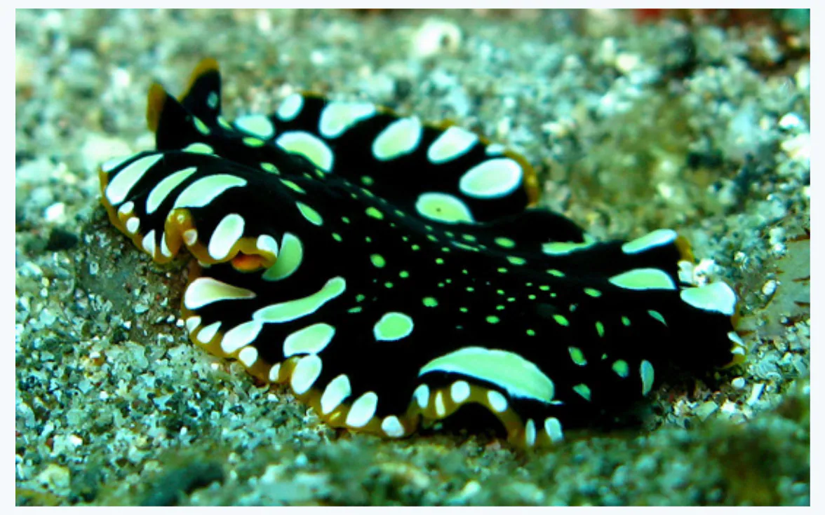
Take the flatworm shown below. It has no lungs or gills. Instead, its broad, flattened body gives it a large surface area relative to its volume. Oxygen simply diffuses across its outer membrane directly into its cells, and carbon dioxide diffuses out the same way. This works because every cell is close enough to the surface that diffusion distances are tiny.
Why can't a large animal like a human rely on direct diffusion for gas exchange?
Tracheal System in Insects
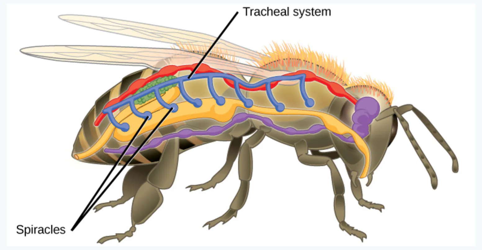
Insects face the same problem — they need oxygen deep inside their bodies — but they solve it differently from mammals. Instead of using the circulatory system to carry oxygen, insects have a tracheal system: a network of branching air tubes that delivers oxygen directly to tissues throughout the body.
Air enters the system through openings called spiracles — small holes along the insect's body wall. From there, the tracheal tubes branch into smaller and smaller passages, reaching every muscle and organ. This direct delivery system is highly efficient for small-bodied animals.
How does the insect tracheal system differ from the mammalian circulatory system in terms of oxygen delivery?
Skin and Gills in Fish
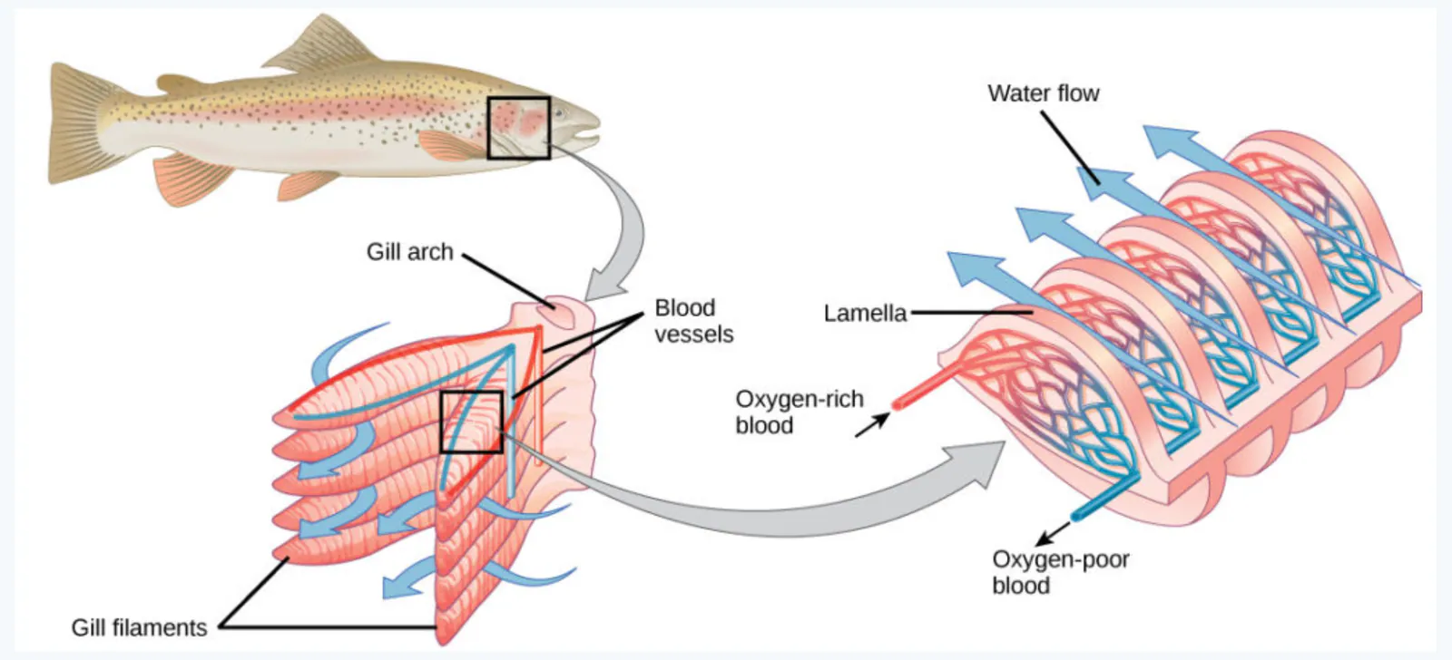
Fish extract oxygen from water, which is much harder than extracting it from air because water contains far less oxygen. They do this using gills — specialized respiratory organs located behind the head.
The structure of a fish gill is beautifully designed for efficiency:
Gill arch: the bony or cartilaginous support structure
Gill filaments: thin, finger-like projections extending from the gill arch, increasing surface area
Lamella (plural: lamellae): tiny, plate-like structures on each gill filament where gas exchange actually occurs
The key innovation is countercurrent exchange: blood flows through the lamellae in the opposite direction to water flowing over the gills. This maintains a concentration gradient along the entire length of the lamella, allowing oxygen to keep diffusing into the blood even after much of it has been absorbed.
Why is countercurrent flow more efficient than having blood and water flow in the same direction?
Mammalian Respiratory System
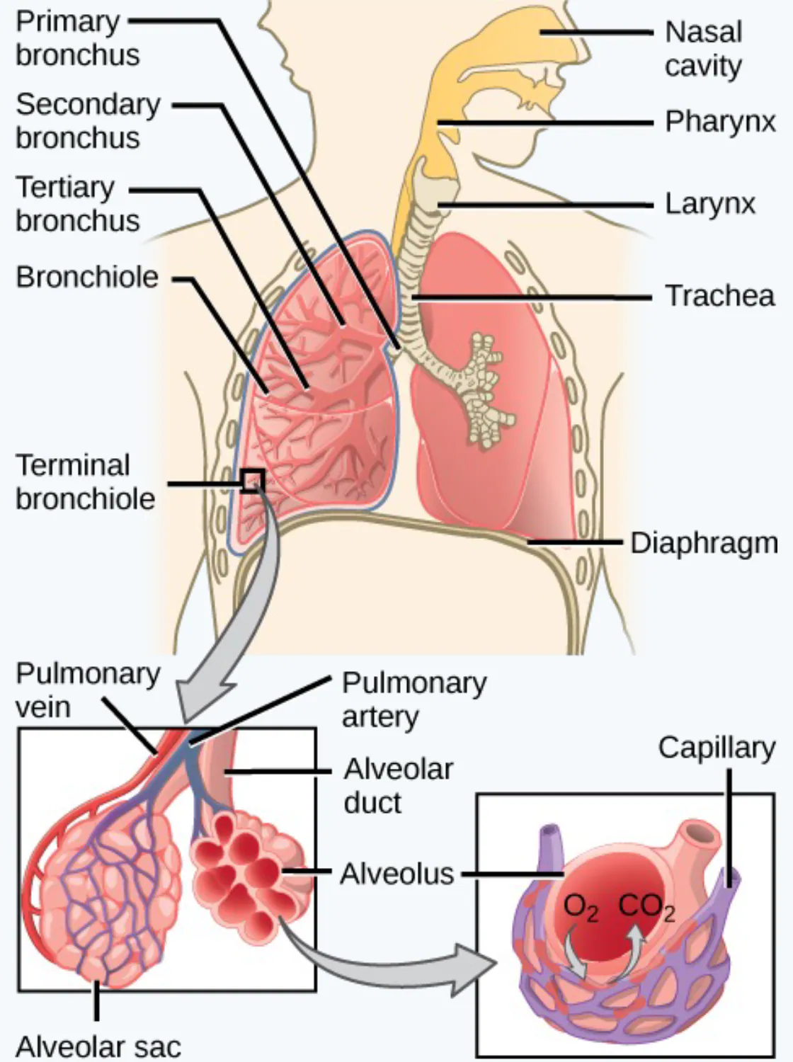
The mammalian respiratory system is a branching network that brings air from the outside environment deep into the lungs, where gas exchange occurs. Here are the major structures, in the order air passes through them:
Nasal cavity: the space inside the nose where air is warmed, moistened, and filtered
Pharynx: the throat region that connects the nasal cavity to the larynx
Larynx: the voice box, which also prevents food from entering the airway
Trachea: the windpipe, a tube that carries air from the larynx toward the lungs
Bronchus (plural: bronchi): the trachea splits into two primary bronchi (one per lung), which branch into secondary and tertiary bronchi
Bronchiole: smaller branches of the tertiary bronchi; the finest bronchioles are called terminal bronchioles
Alveolar duct: a passage leading from the terminal bronchiole to clusters of air sacs
Alveolus (plural: alveoli): tiny, cup-shaped air sacs where gas exchange happens
Alveolar sac: a cluster of multiple alveoli connected by an alveolar duct
Diaphragm: a dome-shaped muscle at the base of the thoracic cavity that drives breathing
Pulmonary artery: carries oxygen-poor blood from the heart to the lungs
Pulmonary vein: carries oxygen-rich blood from the lungs back to the heart
Capillary: tiny blood vessels surrounding the alveoli, where O₂ and CO₂ are exchanged
Trace the path of an oxygen molecule from the nasal cavity to the bloodstream.
Trachea and Bronchi
The trachea is a flexible tube about 10–12 cm long in humans, reinforced with C-shaped cartilage rings that keep it open. It splits into two primary bronchi — one entering each lung. Within each lung, the primary bronchus branches into secondary bronchi (one per lung lobe), then into tertiary bronchi, and finally into bronchioles. This branching pattern is often called the tracheobronchial tree because it looks like an upside-down tree.
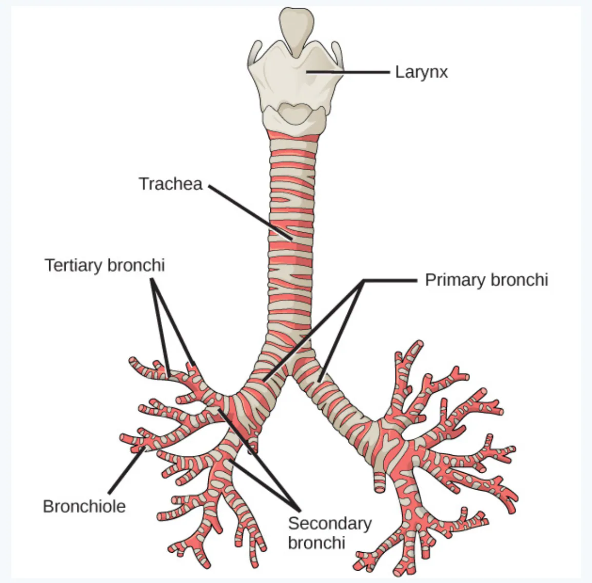
Why does the trachea need cartilage rings?
Lungs
The lungs are the paired organs where gas exchange occurs. The right lung has three lobes (upper, middle, lower), while the left lung has two lobes (upper, lower). The left lung is smaller because it shares space with the heart.
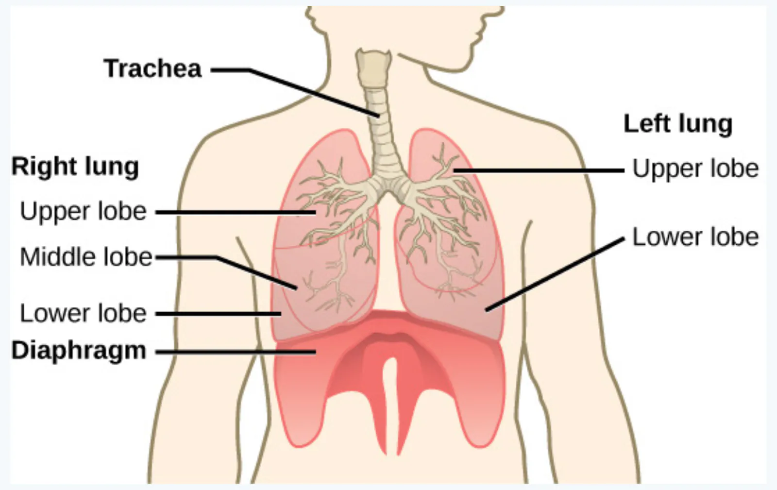
The left lung's smaller size is why the heart sits slightly to the left of center in the chest.
Why does the right lung have three lobes while the left has only two?
Alveoli
The alveoli are the functional units of the lungs — this is where the actual gas exchange happens. Each lung contains about 300 million alveoli, giving a total surface area roughly the size of a tennis court.
Each alveolus is surrounded by a dense network of capillaries. Oxygen (O₂) diffuses from the air inside the alveolus into the blood in the capillaries, while carbon dioxide (CO₂) diffuses in the opposite direction — from the blood into the alveolus, to be exhaled.
The alveolar duct connects the respiratory bronchiole to the alveolar sac, which holds multiple alveoli. The pulmonary artery brings deoxygenated blood to the capillary network, and the pulmonary vein carries oxygenated blood away.
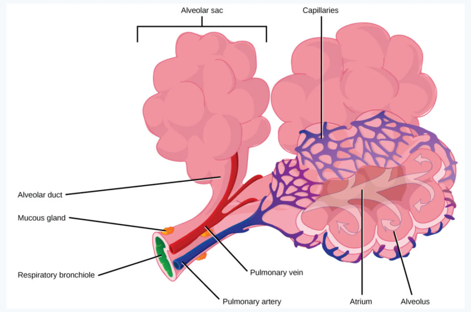
What structural features of alveoli make them efficient for gas exchange?
Protective Mechanisms
The airways need protection from inhaled particles, pathogens, and mucus. The bronchi and bronchioles are lined with cilia — tiny, hair-like projections that beat in coordinated waves. These cilia move mucus (and any trapped particles) upward toward the throat, where it can be swallowed or coughed out. This is often called the mucociliary escalator.
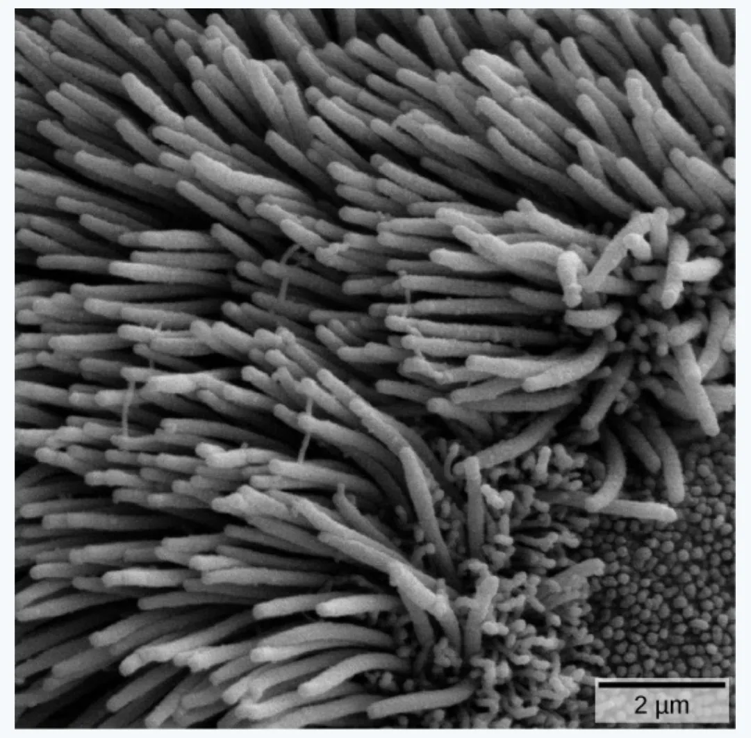
Smoking paralyzes and eventually destroys cilia, which is why smokers develop a "smoker's cough" — without functioning cilia, mucus accumulates and must be expelled by coughing.
What happens to inhaled particles that get trapped in the mucus of the bronchioles?
Lung Capacity
The lungs can hold a large volume of air, but they are not usually filled to maximal capacity. Lung volume measurements are divided into four basic volumes, which can be combined to form capacities.
Lung Volumes
Volume | Definition | Average Value (Adult Male) |
|---|---|---|
Tidal volume (TV) | Amount of air inhaled during a normal breath | 0.5 L |
Expiratory reserve volume (ERV) | Amount of air that can be exhaled after a normal exhalation | 1.2 L |
Inspiratory reserve volume (IRV) | Amount of air that can be further inhaled after a normal inhalation | 3.1 L |
Residual volume (RV) | Air left in the lungs after a forced exhalation | 1.2 L |
Lung Capacities
Capacity | Definition | Equation | Value |
|---|---|---|---|
Vital capacity (VC) | Maximum amount of air moved in or out in one respiratory cycle | VC = ERV + TV + IRV | 4.8 L |
Inspiratory capacity (IC) | Volume that can be inhaled after a normal exhalation | IC = TV + IRV | 3.6 L |
Functional residual capacity (FRC) | Volume remaining after a normal exhalation | FRC = ERV + RV | 2.4 L |
Total lung capacity (TLC) | Total volume after maximal inspiration | TLC = RV + ERV + TV + IRV | 6.0 L |
Forced expiratory volume (FEV1) | Volume forced out in one second | — | ~4.1–5.5 L |
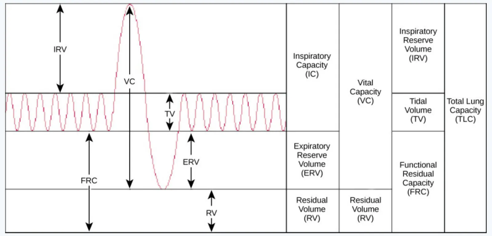
Notice that you can never empty your lungs completely — the residual volume (1.2 L) stays behind. This is why drowning victims can sometimes be revived: there's always some air left in the lungs.
If a person has a tidal volume of 0.5 L and an inspiratory reserve volume of 2.5 L, what is their inspiratory capacity?
Equations for Gas Exchange
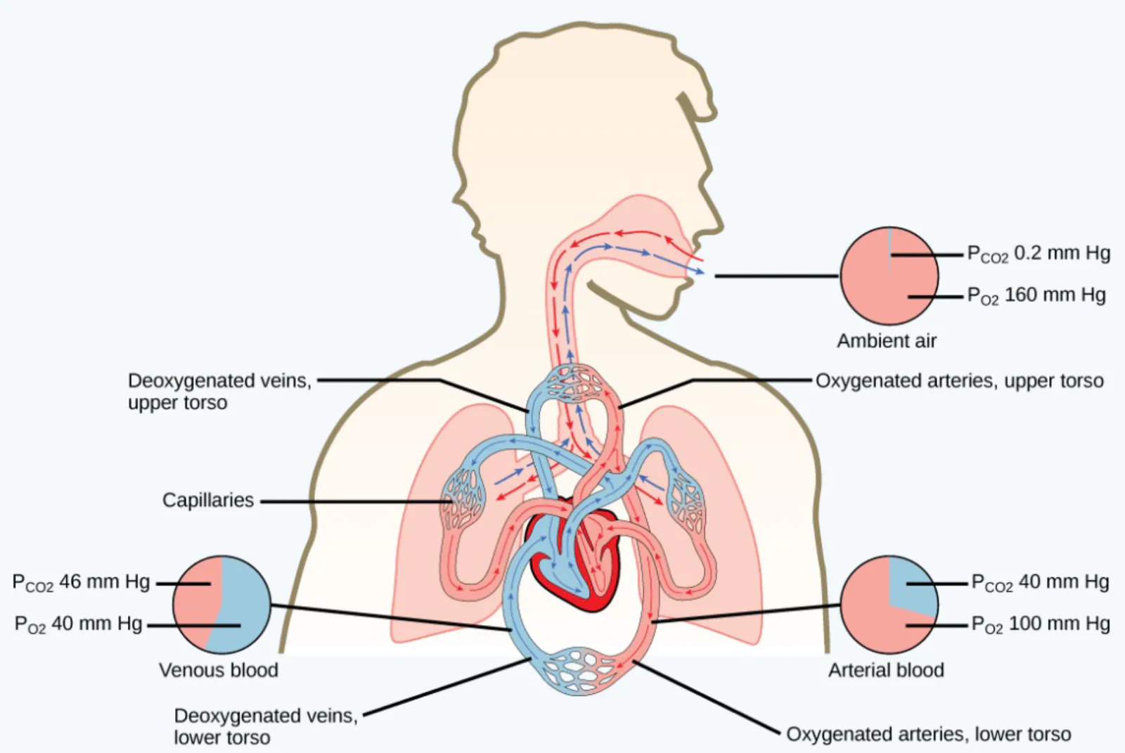
Gases move according to their partial pressures — the pressure exerted by a single gas in a mixture. The partial pressure of a gas equals the total atmospheric pressure multiplied by its percentage in the mixture.
Atmospheric pressure is the sum of all the partial pressures of the atmospheric gases:
For example, since oxygen makes up about 21% of the atmosphere, its partial pressure at sea level is mm Hg.
If carbon dioxide makes up 0.04% of the atmosphere, what is its partial pressure at sea level?
Boyle's Law
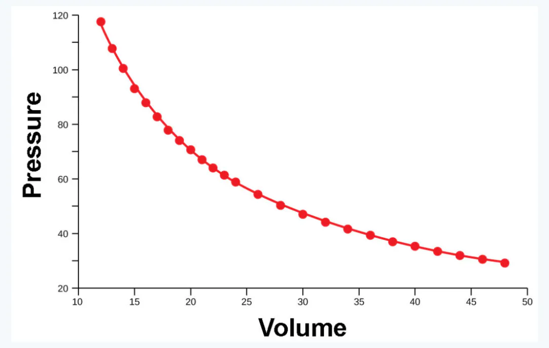
Boyle's Law states that in a closed space, pressure and volume are inversely related. As volume decreases, pressure increases, and vice versa.
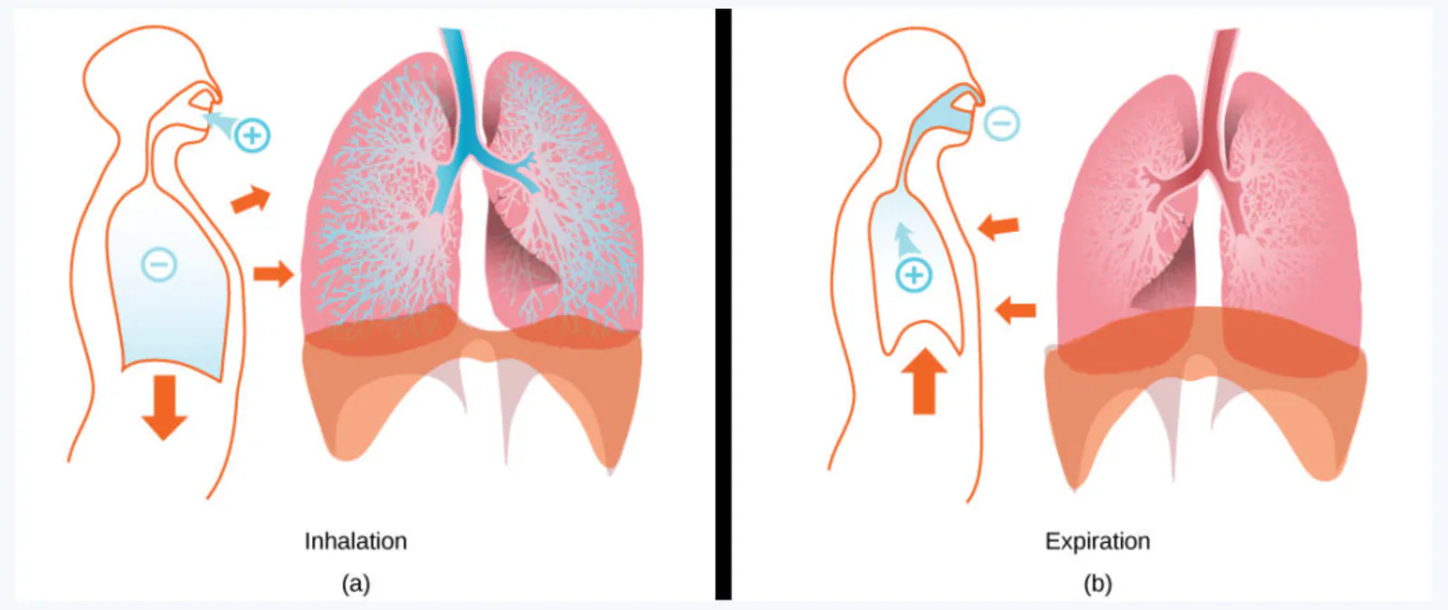
This is the physical principle behind breathing. When the diaphragm contracts and moves downward, the volume of the thoracic cavity increases. According to Boyle's Law, this increase in volume causes the pressure inside the lungs to decrease (below atmospheric pressure), so air rushes in — this is inhalation. When the diaphragm relaxes and moves upward, volume decreases, pressure increases, and air is pushed out — this is expiration.
If you seal a syringe and pull the plunger back, why does the air inside expand? Use Boyle's Law to explain.
The Lungs, Chest Wall, and Diaphragm in Respiration
During inhalation, the rib cage expands outward and the diaphragm contracts and moves downward. This increases thoracic volume, decreases pressure inside the lungs, and air flows in.
During expiration, the rib cage moves inward and the diaphragm relaxes and moves upward. This decreases thoracic volume, increases pressure inside the lungs, and air flows out.
What would happen if the diaphragm were punctured? How would breathing be affected?
Pleura
The lungs are surrounded by a protective tissue layer called the pleura. It has two layers:
Parietal pleura: the outer layer, lining the interior of the thoracic cavity
Visceral pleura: the inner layer, covering the surface of the lungs
Between them is the intrapleural space, a thin, fluid-filled gap. The fluid creates surface tension that holds the two layers together, allowing the lungs to expand and contract smoothly with the chest wall.
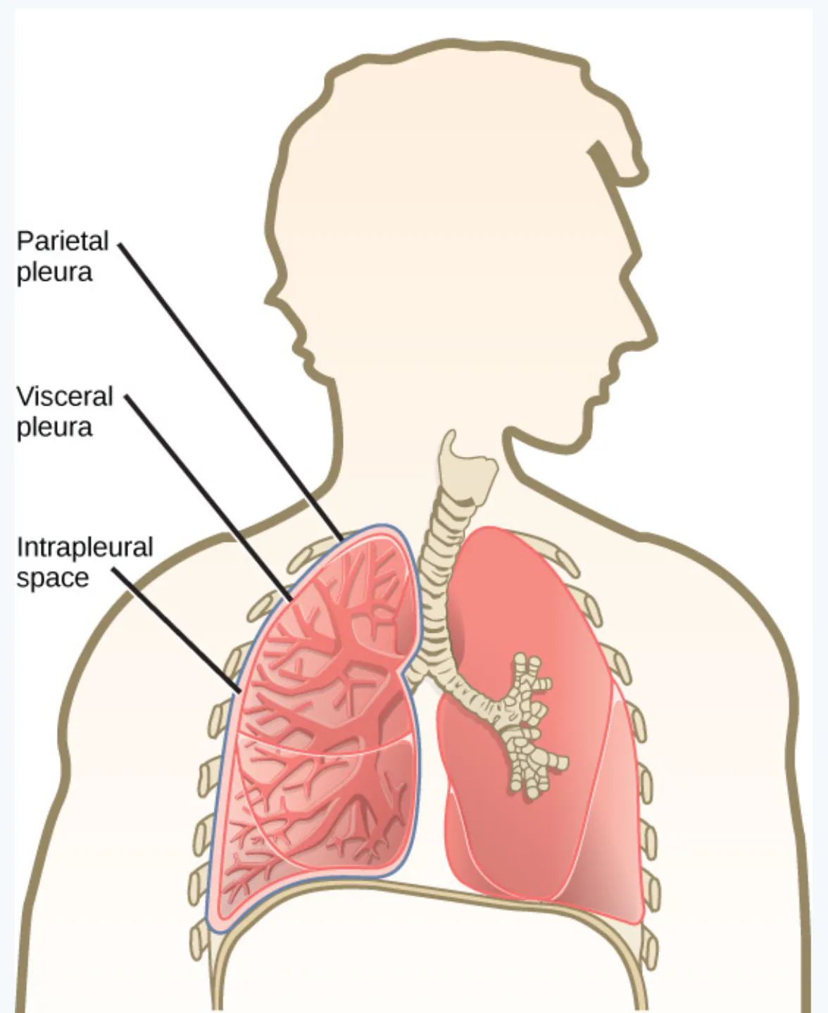
If air enters the intrapleural space (a condition called pneumothorax), the negative pressure is lost and the lung collapses. This can happen from a chest wound or a ruptured lung.
Why is the intrapleural space important for breathing?
The Work of Breathing
Breathing requires work, and this work has two components:
Flow-resistive work: the work needed to overcome resistance in the airways (the alveoli and lung tissues)
Elastic work: the work needed to stretch the intercostal muscles, chest wall, and diaphragm
There is a trade-off: increasing the respiration rate increases flow-resistive work (because air moves faster through the airways, creating more friction) but decreases elastic work (because each breath is shallower, requiring less stretching).
Why does rapid, shallow breathing feel tiring even though each breath is small?
The Ratio of FEV1 to FVC
The FEV1/FVC ratio is a key clinical measurement. FEV1 (forced expiratory volume in 1 second) is the amount of air a person can forcefully exhale in the first second. FVC (forced vital capacity) is the total amount they can exhale.
A healthy person can exhale about 70–80% of their FVC in the first second. This ratio helps distinguish between types of lung disease.
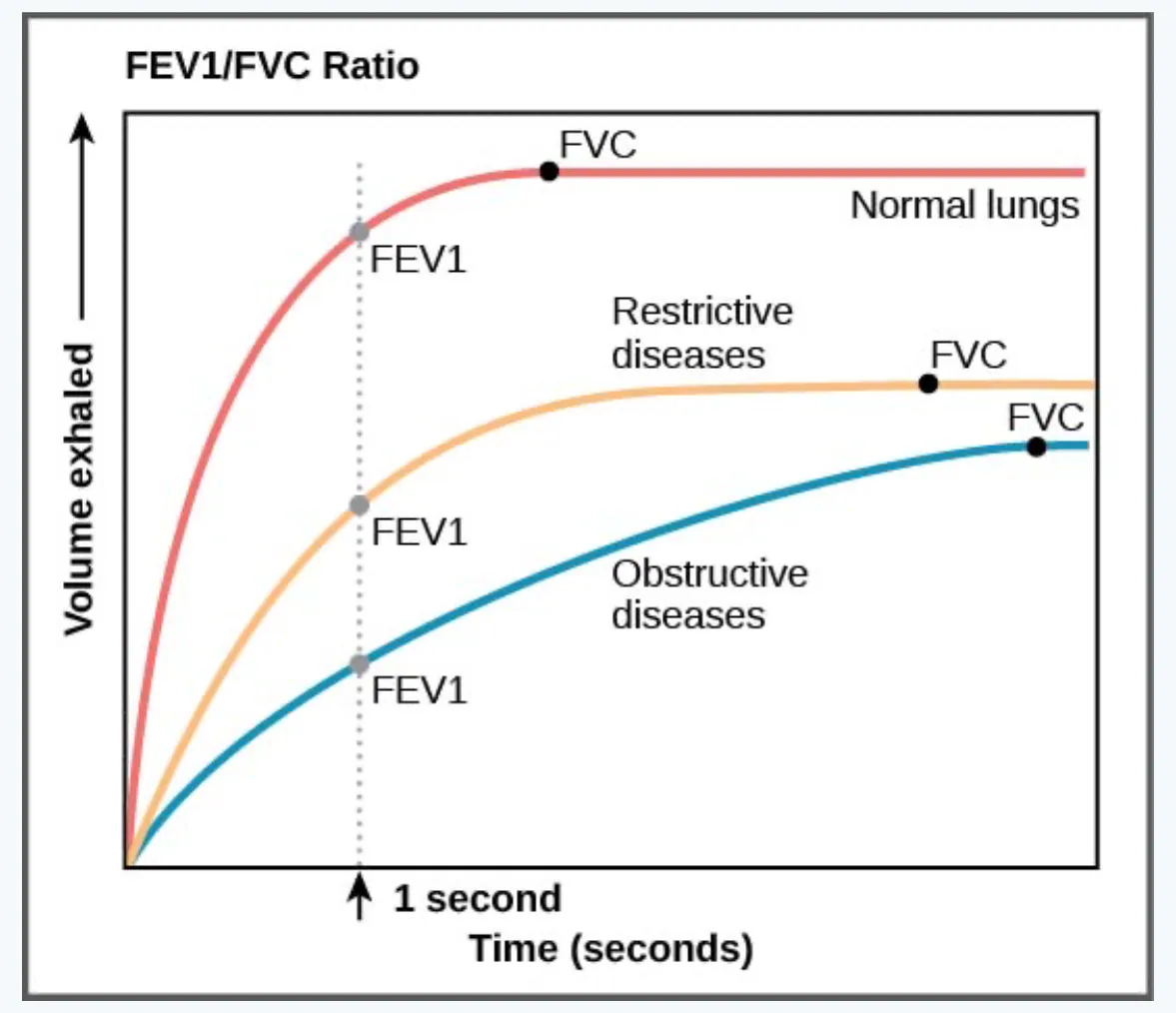
What does a low FEV1/FVC ratio suggest about a person's lungs?
Types of Lung Disease
Lung diseases fall into two main categories:
Restrictive Diseases
In restrictive diseases, the FVC is reduced, but the airways are not obstructed, so the person can still expel air reasonably fast. Examples:
Respiratory distress syndrome: often seen in premature infants whose lungs lack surfactant, causing the alveoli to collapse
Pulmonary fibrosis: scarring of lung tissue that reduces lung elasticity and makes expansion difficult
Obstructive Diseases
In obstructive diseases, airway obstruction results in slow exhalation as well as reduced FVC. Examples:
Emphysema: destruction of alveolar walls, reducing surface area for gas exchange
Asthma: inflammation and narrowing of the airways, often triggered by allergens
Pulmonary edema: fluid accumulation in the lungs, often from heart failure
How would the FEV1/FVC ratio differ between a patient with pulmonary fibrosis and a patient with asthma?
Ventilation/Perfusion Mismatch
Ventilation (V) refers to the amount of air reaching the alveoli. Perfusion (Q) refers to the amount of blood flowing through the pulmonary capillaries.
As cardiac output increases, more capillaries and arteries become perfused (filled with blood). However, sometimes there is a mismatch between ventilation and perfusion — for example, an area of the lung might receive air but not enough blood, or blood but not enough air. This reduces the efficiency of gas exchange.
What happens to blood oxygen levels when a region of the lung is ventilated but not perfused?
Dead Space
Dead space refers to regions of lung tissue that are damaged, blocked, or otherwise unable to participate in gas exchange. Dead space reduces the surface area available for diffusion, decreasing blood oxygen and increasing carbon dioxide levels.
Dead space occurs when no ventilation and/or perfusion takes place:
Anatomical dead space (or anatomical shunt): arises from an anatomical failure — for example, a structural defect in the airways
Physiological dead space (or physiological shunt): arises from a functional impairment of the lung or arteries — for example, a blood clot blocking a pulmonary artery
What is the difference between anatomical and physiological dead space?
Hemoglobin
Hemoglobin is a protein found in red blood cells that carries oxygen. It is composed of two alpha and two beta subunits that surround an iron-containing heme group. Oxygen readily binds to this heme group — each hemoglobin molecule can carry up to four oxygen molecules.
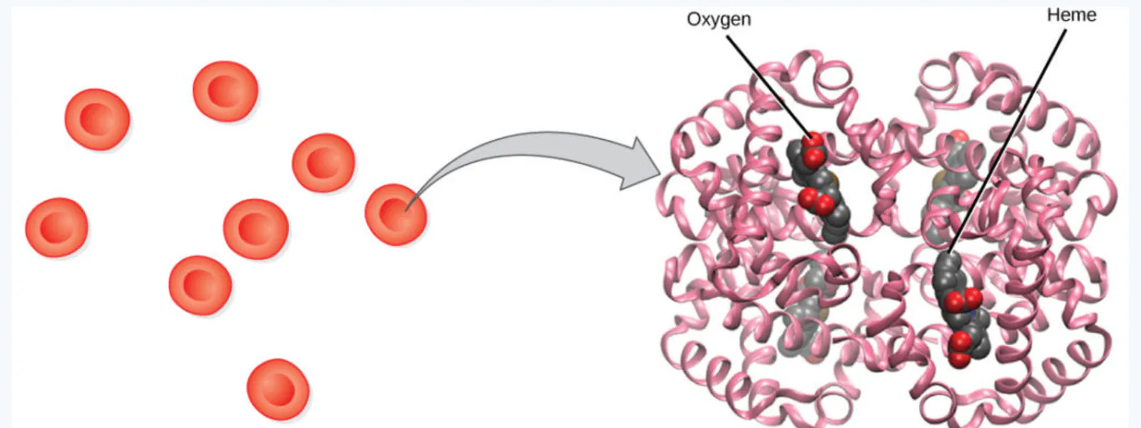
The "heme" in hemoglobin is what gives blood its red color. When oxygen is bound, blood is bright red; when oxygen is released, blood is darker, bluish-red.
How many oxygen molecules can a single hemoglobin molecule carry?
Oxygen Dissociation Curve
The oxygen dissociation curve shows the relationship between the partial pressure of oxygen (PO₂) and hemoglobin saturation. As PO₂ increases, more oxygen binds to hemoglobin — but the relationship is not linear. It is sigmoidal (S-shaped), meaning that hemoglobin loads oxygen readily at high PO₂ (in the lungs) and unloads it readily at low PO₂ (in the tissues).
The curve can shift depending on conditions:
Left shift (higher Hb-O₂ affinity): occurs with lower CO₂, higher pH, and lower temperature. Hemoglobin holds oxygen more tightly.
Right shift (reduced Hb-O₂ affinity): occurs with higher CO₂, lower pH, and higher temperature. Hemoglobin releases oxygen more easily — useful when tissues are metabolically active.
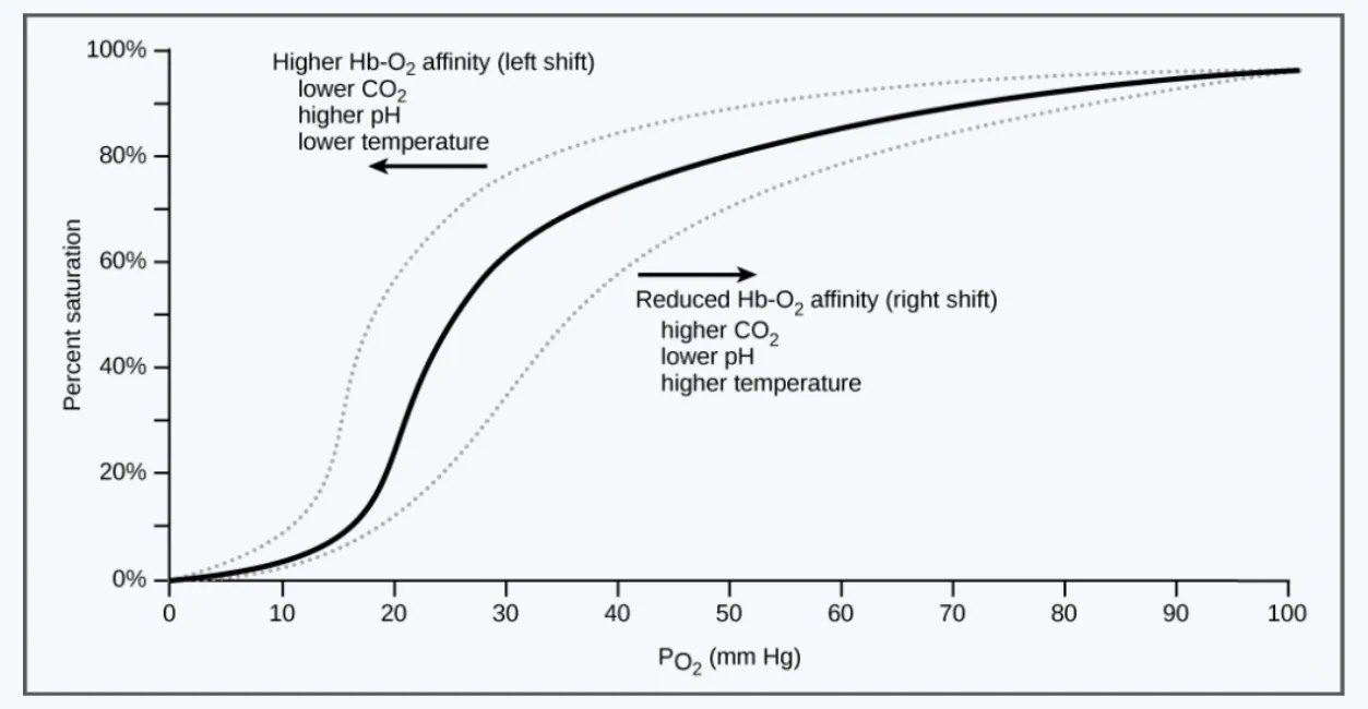
During exercise, muscles produce more CO₂ and heat. How would this affect the oxygen dissociation curve, and why is this beneficial?
Diseases and Oxygen Binding
Sickle cell anemia is a genetic disorder in which red blood cells become crescent-shaped (sickle-shaped) instead of round. These misshapen cells can clog capillaries and break down easily. The abnormal hemoglobin in sickle cell disease has a reduced ability to bind and release oxygen normally.
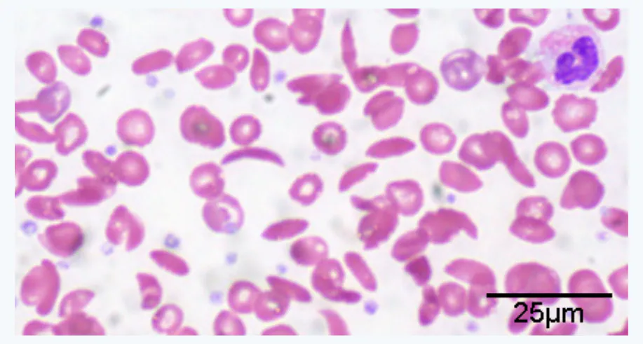
Why would sickle-shaped red blood cells have trouble delivering oxygen to tissues?
Transport of Carbon Dioxide in the Blood
Carbon dioxide is transported from the tissues to the lungs through three methods:
Dissolved directly in the blood — a small amount (about 5–10%)
Bound to plasma proteins or hemoglobin — about 20–25%
Converted into bicarbonate — the majority (about 70%). In red blood cells, an enzyme called carbonic anhydrase converts CO₂ and water into carbonic acid, which quickly dissociates into bicarbonate and hydrogen ions
What is the most common way carbon dioxide is transported in the blood?
Key Takeaways
Simple organisms like flatworms rely on direct diffusion across their body surface
Insects use a tracheal system with spiracles to deliver oxygen directly to tissues
Fish use gills with lamellae and countercurrent exchange for efficient oxygen extraction from water
The mammalian respiratory system is a branching network: nasal cavity → pharynx → larynx → trachea → bronchi → bronchioles → alveolar ducts → alveoli (where gas exchange occurs)
Boyle's Law explains how the diaphragm creates pressure changes that drive inhalation and expiration
Lung volumes (TV, ERV, IRV, RV) combine to form capacities (VC, IC, FRC, TLC), and the FEV1/FVC ratio helps diagnose lung disease
Hemoglobin in red blood cells carries oxygen, and its binding affinity is shown by the oxygen dissociation curve
Carbon dioxide is transported mainly as bicarbonate in the blood
अब यही अपने लेक्चर के लिए सोचकर देखें।
अपना पहला regra मुफ़्त में तैयार करेंआपके पहले लेक्चर मुफ़्त हैं, हर फ़ीचर के साथ। कार्ड की ज़रूरत नहीं।
स्रोत स्लाइड Shelli Carter और Lumen Learning की “Biology for Majors II” से ली गई हैं, जिनमें OpenStax Biology (cnx.org) की सामग्री शामिल है; लाइसेंस: CC BY 4.0